
Lungs
last authored:
last reviewed:
Introduction
The lungs are located in the thorax. The right lung has 3 lobes, 55% of total lung, while the left has two lobes. Each weighs x-x. and is x large. They are covered with the thin pleural lining - only a few cells thick. Between the lungs and chest wall is the pleural space, a very small (potential space) which contains fluid to lubricate the lungs.
Lungs are extremely elastic due to the rigid bony thorax and their cellular and connective composite.
Anatomical Landmarks
lung landmarks
- inferior margins: 6th (anterior)
8th (lateral), and 10th (posteriorly) ribs - oblique fissures: diagonal, from T3 to the 6th rib anteriorly
- minor fissure: horizonal line from anterior 4th rib
- pleura is at 8,10, 12
- major fissure goes to T3/T4 posteriorly
The majority of the lung is composed of acinar sacs, leading to a surface area of 50-100 m2 per lung. The acinar sacs contain 300 million gas-exchanging alveoli.
Distal Airways
Bronchioles are bifurcations that do not contain cartilage, making them collapsible. They are also contractile due to smooth muscle. This is a principal location of asthma reactivity.
Lobules contain clusters of alveolar sacs attached to terminal bronchioles. These sacs, or acini (one acinus), are composed of respiratory bronchioles and alveoli and are the true site of ventilation.
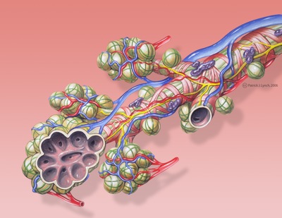
Bronchial anatomy, Patrick Lynch and Carl Jaffe (license)
Alveoli are gas-exchanging structures. They begin as outpouches of respiratory bronchioles but are found in greatest number in alveolar sacs.
Alveoli are in intimate contact with the endothelial cells of the lung's extensive capillary system.
The thin Type I pneumocytes make up most of the alveolar walls, while type II pneumocytes (5% if the cells) secrete surfactant to reduce surface tension and keep the alveoli inflated. Type II pneumocytes are capable of regeneration and differention into type I cells following injury.
Macrophages live in alveoli.
Cellular Composition
Between acinar sacs is the interstitium, which consists of the basement membrane of alveolar and endothelial cells - fused in the thinnest regions - and connective tissue in between. This includes:
- collagen fibres (structural)
- elastic fibres (elastic)
- proteoglycans (hydration and open space)
- fibroblasts (extracellular matrix producers)
- mast cells (innate immunity cells)
- occasional lymphocytes and monocytes (adaptive and innate immunity cells)
Resources and References
 |