
The Kidneys
last authored: Jan 2010, David LaPierre
last reviewed:
Introduction
Kidneys are exquisitely verstatile organs, regulating blood volume and osmolarity, filtering wastes, and controlling calcium and phosphate balance. Together, the kidneys filter about 180L of plasma per day, or 125 ml/min. Kidneys are located retroperitoneally in the lower thoracic/upper lumbar region. Each kidney weighs 120-170 g.
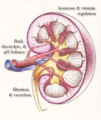
Kidneys have two distinct regions - the pale cortex and the dark inner medulla, with accompanying 6-15 pyramids. The hilum contains vasculature, lymphatics, nerves, and the renal pelvis.
The kidneys receive about 20% of total cardiac output through the renal arteries, which leave the aorta at the first lumbar vertebra. Renal arteries branch many times to end in afferent arterioles. Efferent arterioles leave the glomerulus and travel alongside the tubules to provide fluid and solute exchange.
The kidneys are richly innervated by sympathetic nerve endings, across both the vasculature and the parenchyma. They increase renin secretion from juxtaglomerular cells.
The Nephron
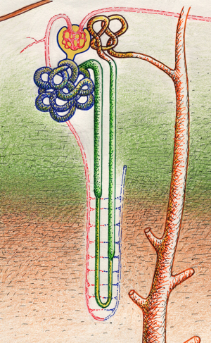
Each kidney contains 800,000 to 1,200,000 nephrons, the basic functional unit of the kidney. Each is composed of two main components: the glomerulus and its attached tubule.
Nephrons are classified according to length of loop of Henle. Long loops originate from the corticomedullary region and extend deep into the inner medulla, while short loops (most common) begin in the superficial and midcortical regions and extend to the outer medulla.
The proximal tubule is continuous with Bowman's capsule and consists of two segments - the proximal convoluted tubule in the cortex and the straight proximal tubule in the medulla.
The thin descending loop of the Loop of Henle dips in the medulla to give rise to the thick ascending loop and distal convoluted tubule.
The connecting segment gives rise to the collecting segment, which includes the cortical collecting ducts and medullary collecting ducts. These structures differ in their cell types, transporters, and hormine responsiveness. The collecting ducts terminate in the papillary collecting ducts, or ducts of Bellini, which empty urine into the renal pelvis.
The Glomerulus
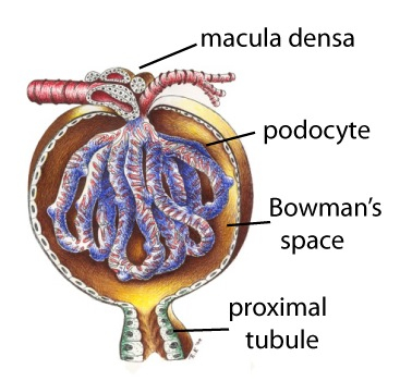
The glomerulus is a unique network of lobular capillary tufts that is fed by afferent arterioles and drained by efferent arterioles. The entire glomerulus is enclosed within Bowman's capsule, an epithelial structure. The amount filtered is only about 20% of renal plasma flow (filtration fraction = 0.20).
The core of the glomerulus contains smooth muscle-like mesangeal cells and the extracellular matrix, rich in fibronectin, they produce. This provides a framework for the capillaries, and, due to its contractile properties, can control blood through through the nephron. Abnormal matrix production can occur in many forms of glomerular disease.
Fluid flowing from the capillary has three structures to cross: the capillary, through the basement membrane, and through the filtration slits of podocytes.
Endothelial Cells
The glomerular capillaries are lined by endothelial cells with fenestrae of about 70nm that allow for a high ultrafiltration coefficient. These cells are covered with a coat rich in polyanionic glycoproteins, reducing permeability of negatively charged proteins.
Endothelial cells regulate coagulation, inflammation, and vascular tone.
Podocytes
This capillary bed is covered by epithelial cells called podocytes which extend many foot-like projections to provide a filtration net. Fluid flows through 10 nm fenestrae, or slit diaphragms, and through the glomerular basement membrane before encountering epithelial podocytes. Damage to these podocytes can result in proteinuria, present in a the majority of kidney diseases.
Basement Membrane
The basement membrane lies between the endothelial and epithelial cells. Both cell types form a layer of hydrated gel, comprised of glycoproteins and interwoven collagens IV and V . It contains negative charges in sialiac and carboxylic acids to prevent flow of proteins and othe rmolecules greater than 1 kDa.
Juxtaglomerular Apparatus
Next the site of arteriole entry and exit, distal tubule cells are tightly adherent to the glomerulus and make up the macula densa of the juxtaglomerular apparatus. These cells regulate glomerular filtration and renin formation in response to NaCl concentration and sympathetic innervation.
Filtration
The glomerulus forms an impressive filtration barrier using molecular size, shape, and charge.
Additional Resources
http://www.wramc.amedd.army.mil/education/lecture/nephrology/Pages/default.aspx
http://www.gamewood.net/rnet/renalpath/ch3.ht
http://www.duj.com/
http://www.urologychannel.com/
 |