
The Cardiovascular System
last authored: Jan 2010, David LaPierre
last reviewed:
Introduction
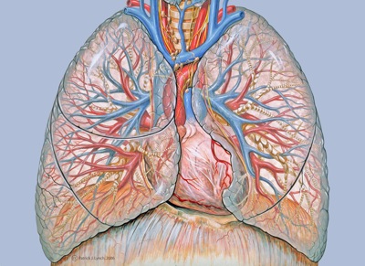
used with permission, Patrick Lynch
Central in the body and to life, the cardiovascular system pumps blood around the body to provide it with a steady supply of oxygen, energy, and nutrients while removing wastes.
The heart is the central player of the cardiovascular system, pumping blood along the almost 100,000 km of blood vessels. Blood pressure is the force within the vasculature, which includes arteries, veins, and capillaries.
The Heart
- overview
- vessels and chambers
- the heart beat
Overview
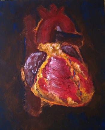
used with permission, denn
The heart is a muscular, fist-sized organ located in the centre of the chest and weighing 250-350 g. It is a remarkably effecient and reliable pump, propelling over 6000 litres of blood daily and beating more than 40 million times yearly, or over 3 billion times over a lifetime.
The heart lies centrally within the chest, at the levels of the x and x ribs.
The heart is contained in the pericardium, a tough, membranous sac bounded by the pleural sacs of the lungs and the diaphragm below. The pericardial cavity is filled with pericardial fluid, lubricating the heart and allowing it to beat smoothly.
The heart consists of three layers: the epicardium, a thin layer of epicardial and connective tissue; the myocardium, a thick layer of contractile cardiomyocytes, and the thin endocardium, composed of endothelial cells. Myocytes comprise only 25% of the number of heart cells but account for almost 90% of its volume. The remainder are mostly endothelial cells of the capillary network and fibroblasts. Extracellular matrix and leukocytes are rare.
The heart criticially depends on oxygen for its function. Altough it counts for only 1/200 of the body's weight, it requires almost 1/20 of total blood supply at rest. It therefore has a rich vasculature to keep it perfused - the coronary (Latin: crown) arteries, 5- to 10 cm long and 2-4 mm in diameter. Atherosclerosis of coronary vessels is the major cause of angina and the acute coronary syndromes.
Vessels and Chambers
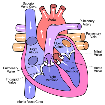
Blood returning from the body through the inferior and superior vena cavae enter the right atrium and pass through the tricuspid valve to enter the right ventricle.
From there, blood is pumped through the pulmonary valve to the lungs, where blood is oxygenated.
Blood returns to the left atrium and passes through the mitral valve into the muscular left ventricle, where is pumped across the aortic valve and into systemic circulation.
The heart is four chambered, and while they perform essentially different roles in the body, they work completely with each other. Throughout the cardiac cycle, the right and left atria fill with blood coming from the body and lungs, respectively.
The Heart Beat
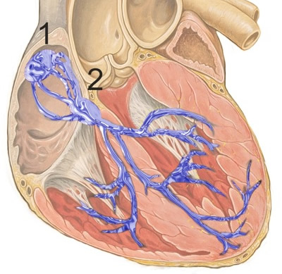
Embedded within the heart is an electrical conducting system, facilitating the coordinated contraction of the cardiac cycle.
The heart rate and thereby cardiac output are controlled by internal pacemaker systems and external neural and endocrine systems, achieving its ultimate goal of maintaining appropriate blood pressure.
Blood Vessels
- overview
- arteries
- veins
- capillaries
Overview
The braching network of blood vessels carries blood from the heart to the tissues of the body. End-to-end, the body's blood vessels would stretch almost 100,000 km.
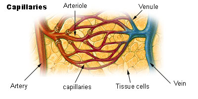
Arteries transport blood from the heart to capillaries, where nutrient and waste diffusion occurs. Veins cary blood back to the heart.
Endothelial cells line the inside of the cardiovascular system. Regulation of CV function is mediated primarily by smooth muscle cells and cardiomyocytes.
The enormous branching of the vasculature creates large differences in cross-sectioanal area and flow.
Vessels |
Number |
Aggregate cross-sectional area (cm2) |
Mean velocity (cm/s) |
|---|---|---|---|
Aorta |
1 |
4 |
21 |
Arteries |
8000 |
63 |
1.3 |
Arteriloles |
2 x 10⁷ |
141 |
0.6 |
Capillaries |
1-4 x 10¹⁰ |
2827 |
0.03 |
Blood flows through these systems due to differences in pressure at either end.
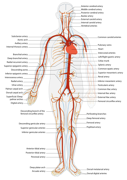
Arteries
Arteries are the thick-walled vessels transporting blood from the heart to tissue beds. Their stiff but elastic walls allow them to stretch under proessure and provide constant blood pressure to capillaries .The aorta is roughly 2.5 cm in diameter, while arterioles have a diameter of 0.3 mm or less.
Microvascular control ensures blood gets to the right spots. The sympathetic system can shut down blood flow to skeletal muscle and the skin in order to pool blood in the vital organs. Precapillary sphincters serve as gates to control blood flow into individual capillary beds.
see pg 478, B and B, to learn about local regulation of vessels using O2, CO2, pH, lactic acid, and others.
used with permission, Mariana Ruiz Villarreal
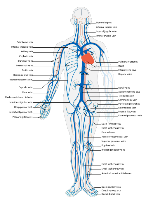
Veins
Veins contain most of the systemic circulation's blood volume due to their large compliance, giving them the name 'capacitance vessels'. This is not due so much to the elastic properties of their walls, but rather their ability to change shape.
used with permission, Mariana Ruiz Villarreal
Capillaries
Capillaries are the smalles of vessels, made up simply of porous endothelial cells. It is here that gases, nutrients, and wastes diffuse between the cardiovascular system and the tissues.
Blood Flow
Most (85%) of the body's 5L of blood is in the systemic circulation, with 10% in the pulmonary circulation and 5% in the heart. 65% of total blood is in the systemic veins.
Hemodynamics describes blood flow throughout the vessels of cardiovascular system.
With a stroke volume of ~ 70 ml, and heart rate of 70 bpm, mean aggregate flow throughout the CV system is 80 ml/s. This value doesn't change as the cross sectional area grows from 4 cm2 for the aorta to over 2800 cm2 for the capillaries, which means that velocity in the capillaries decreases quite a lot.
The largest cross sectional area is in the post-capillary venules, however.
Alterations in normal fluid homeostasis results in edema.
Heart Output |
5000 ml/min |
skeletal muscle |
1450 |
GI system |
1250 |
kidney |
1000 |
brain |
700 |
skin |
375 |
coronary circulation |
225 |
The cardiovascular system has close relationships with:
- the respiratory system, to provide oxygenated blood to the body and remove gaseous wastes.
- the kidneys, regulating blood pressure and fluid levels to maintain homeostasis.
- the autonomic nervous system and endocrine system, which regulate cardiac output and perfusion.
Development of the cardiovascular system is important and interesting.
Resources and References
University of Utah WebPath images of the heart.
Canadian Cardiovascular Society
American Cardiology Society
 |