
Chest X Ray
last authored: Oct 2010, David LaPierre
last reviewed:
Introduction
The Chest X-Ray (CXR) is the most common imaging modality done due to its ability to quickly and easily provide much information. It can eliminate some structural abnormalities from consideration and is also sufficient, along with history and physical exam, to make many diagnoses.
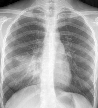
Right middle lobe pneumonia, courtesy of ChestAtlas.com
Differentiation of structures is possible because of differing densities. There four basic densities include: air (aerated lung, trachea), fat, soft tissue (heart, liver, diaphragm), and bone.
Structures of differing densities in contact with each other will be discernable, while tissues of smiliar density will not.
Approach to the CXR
- technique
- surrounding
structures - mediastinum
- lungs
- pulmonary
findings - cardiac
findings
Technique
The first step of CXR, or any diagnostic test, is to assess for technical adequacy.
Patient identification and date of image: ensure you're examining the right film!
Projection: ideally, a CXR will be taken PA and lateral views, with the patient upright. Portable studies tend to be AP, which are less instructive. Specifically, AP views magnify thoracic structures and provide less resolution.
Patient position:Ideally, the patient is upright, with arms extended. If not, position should be noted. Another important variable is rotation. To assess, compare the distances between the spinous processes and the clavicular heads. Lastly, assess lordosis. It is best if the coronal planes of both patient and film are perpindicular to X-ray path.
Adequacy of view: Ensure the entire lungs are visible - costophrenic angles, apices, and sides, on both PA and lateral views.
Penetration: Level of exposure is very important. Overpenetration will cause structures to be more radiolucent (black) than reality, while underpenetration will cause the opposite. It is critical to compare dfferences in penetration between CXRs taken at different times.
To quickly assess penetration, evaluate the vertebral bodies. They should be well visualized through the neck and behind the trachea, faintly visualized through the heart, and not at all through the diaphragm.
Inspiration/expiration: Unless noted the CXR should be taken during maximal inspiration, meaning at least 10 posterior rib shadows are visible over the lung fields.
Expiratory views accentuate pulmonary vasculature and cause the heart shadow to appear elevate or enlarged, falsely suggesting pulmonary edema or cardiomegaly.
Structures
When evaluating a CXR, the lungs are the last structure you should look at. For each CXR, look in the following sequence:
Abdomen: The liver is always visible, as is, often, the spleen. Gas-containing structures include the gas bubble of the stomach and the hepatic and splenic flextures of the colon. Free air under the diaphragm suggests a perforated viscus and should be looked for on every slide.
Diaphragm: The right is often higher than the left. Abnormal elevation of a hemidiaphragm can be due to volume loss (atalectasis), as a result of fluid collection, or because of phrenic nerve paralysis.
Soft Tissues: assess for abnormal densities suggestive of a mass or fluid collection.
Ribs: Follow the contours of each rib, which should be smooth and uninterrupted. Note any swelling (healing fracture or hematoma) or fracture. Focal loss suggests lytic destruction, as can occur with malignancy.
Shoulder girdle: Look for dislocations, fractures, or lytic lesions.
Mediastinum
Inspect for focal or diffuse widening or the presence of air. In general, the mediastinum can be divided into three compartments on lateral view. Draw a line anterior to the trachea but posterior to the heart. This divides the anterior and middle compartments. A line 1 cm in front of the vertebral bodies denotes the posterior compartment.
Anterior compartment: This contains the heart and retrosternal clear space. Abnormal masses are most often caused by the "Five T's":
- thyroid masses
- thymomas
- teratomas
- thoracic aortic aneurysms
- terrible lymphomas
Middle compartment: This contains the esophagus, trachea, aortic arch, and lymph nodes. Enlarged nodes are the most common cause of middle mediastinal masses. Look at the trachea and esophagus for signs of narrowing, compression, or deviation.
Posterior compartment: This contains the vertebral bodies, descending thoracic aorta, spinal nerves, and lymph nodes. On lateral view, inspect vertebral bodies for alignment, disk space enlargement or narrowing, fractures, or lytic lesions.
Lungs
Pleura: There are two pleural layers, separated by a potential space. Look for the presence of thickening, masses, pneumothorax, or pleural effusions. Assess the costophrenic angle for effusions, which are best viewed on lateral view.
Lungs: Examine each lung in turn, then compre with the contralateral lung. Compare with an older CXR if at all possible.
The lungs are divided by fissures into lobes - three on the right, and two on the left. Both frontal and lateral views are helpful in localizing lesions.
Have picture here of lung lobes
CXR Findings
Most lung diseases cause a increase in radiodensity of the lung.
It can be helpful to use the following descriptors:
- focal vs diffuse
- homogenous vs heterogenous
- well circumscribed vs poorly defined
- size (<3cm = nodule; >3cm = mass)
- linear/reticular vs nodular
- acute vs chronic
- improving vs worsening
Interstitial disease: The interstitium contains supporting structures including pulmonary vessels, bronchi, and connective tissue. On normal CXR, the visible interstitium consists of the pulmonary vessels, which are most pronounced at the hila and decrease in prominence towards the periphery.
If increased radiodensity is due to the interstitium, its structures will appear more pronounced. If generalized or diffuse, a linear or reticular pattern will be visible, while if localized, multiple tine nodules may be present. Common causes include pulmonary edema, inflammation, fibrosis, and tumour.
Airspace disease: Alveoli are filled with air and accordingly appear dark on CXR. If these airspaces fill, with fluid, blood, pus, or cells, they will appear more radiodense. This pattern can be relatively homogenous or patchy and diffuse. Airspace disease commonly is accompanied by the Silhoutte and air bronchogram signs, described below.
Silhouette sign: Loss of normal radiographic contours of the heart in the setting of lung disease. It is usually found in conditions resulting in airspace disease, rather than interstitial disease. Complete consolidation of a given lobe will produce characteristic silhouette signs:
- RUL: loss of upper right heart border, asending airta
- RML: loss of right heart border
- RLL: loss of right hemidiaphragm
- LUL: loss of aoric knuckle, left atrium
- LLL: loss of left hemidiaphragm
- lingula: loss of left heart border
Air bronchogram sign: Normally the trachea and proximal bronchi are visible because they represent air-filled structures next to the soft-tissue of the mediastinum. Smaller bonchi are normally not visible because they are in contact with the air-filled lung.
If, however, normal, aerated lung is replaced by tissue increased density, as can occur in consolidation, the smaller airways become visible and form air bronchograms. However, if bronchi themselves are also filled with fluid, air bronchograms will not be present.
Cardiac Findings
The CXR provides much information regarding structure and function of the heart and great vessels.
Cardiac enlargement is present when the cardiac silhouette is greater than 1/2 the diameter of the thorax. The heart may be falsely enlarged due to poor inflation, and with an AP view.
Left atrial enlargement is suggested when the left-sided heart border is straightened or bulges to the left. The main bronchi may be widely splayed, and a circular opacity or double density may be present.
Right atrial enlargement is suggested by a right-sided bulge.
Left ventricular enlargement results in postereo-lateral displacement of the apex, with rounding suggesting hypertrophy.
Right ventricular enlargement is best seen on lateral view and may be present when the right ventricular border occupies more than 1/3 of the retrosternal space between the diaphragm and the thoracic apex.
The aorta can become dilated with severe atherosclerosis, hypertension, or dissection.
Pulmonary hypertension or increased vascular resisitance can result in dilation of the proximal pulmonary vessels.
Heart failure, with attending increased left heart pressure, leads to pulmonary venous congestion, redistribution of blood flow to the lung apices, and transudation of fluid into the interstitial space along fissures and the lung's lower margins (Kerley's B lines). Further increases results in fluid collection in the airspaces, normally in the inner 2/3 of the lung fields early on to yield characteristic butterfly appearance.
Calcification can occur in the pericardium, coronary arteries, or valves.
Resources and References
Queen's University 'Approach to CXR'
ChestAtlas.com - open access database of CXR
Topic Development
authors: DLP, Aug 09
editors:
reviewers:
 |