
Gram Stain
last authored: Feb 2010, David LaPierre
last reviewed:
Introduction
The Gram stain allows differentiation of most bacteria into two groups corresponding to cell wall type. Gram positive cells contain thick peptidoglycan with numerous techoic-acid cross-linkages which bind to the highly basic Crystal violet dye.
- sample
preparation - staining
protocol - microscopic
evaluation
Sample Preparation
- sputum
- blood
- pus
- urine
Sputum
Sputum should be free of squamous epithelial cells (<5 per high-power field)and should contain neutrophils (10-15 per field) to confirm it is from the lower respiratory tract and is not contaminated with oropharyngeal secretions.
If possible, rinse mouth before collection. Cough deeply and spit directly into a sterile cup, avoiding contact with saliva. Bronchoscopy specimens may be used, though anesthetics can inhibit bacterial growth.
Most lower respiratory tract pathogens grow within 2-3 days.
Staining Protocol
Fix bacteria and cells on the slide with 95% methanol for 1 minute, OR with heat. Allow methanol to run off as the slide airdries.
Apply the primary crystal violet stain for 10-30 seconds without allowing the slide to dry. Rinse with tap water, shaking off the excess.
Flood the slide with iodine to fix the crystal violet to the bacterial cell wall. Keep the iodine on the slide for twice as long as the crystal violet.
Flood the slide with decolorizer (alcohol/acetone) to remove the stain from gram negative bacteria. Flood for 10 seconds and immediately rinse with tap water. Repeat until blue dye no longer runs off the slide.
Flood the slide with safranin counterstain for 30 seconds. Rinse with tap water and pat dry or airdry.
Gram-stained specimens should be examined using various lenses up to x1000 (oil-immersion.
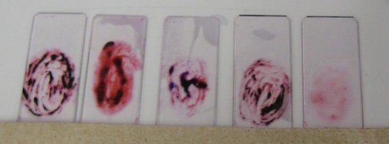
Gram-stained sputum smears
Presence of bacterial cells, Gram reactions, morphologies, and arrangements, all provide diagnostic information.
The presence of a predominant organism, particularly if found within leukocytes, suggests it is the likely pathogen.
If inflammatory cells but no pathogens are seen, a large number of possibilities exist, many of which are nonbacterial.
 |