
Pneumonia
last authored: Feb 2010, David LaPierre
last reviewed:
Introduction
Pneumonia describes various infections of the lower respiratory tract. It is an important cause of death, especially during adult years, but is readily reversible. Every physician should know how to recognize and treat pneumonia.
Causes and Risk Factors
Risk factors for pneumonia include:
- smoking, toxic inhalation, aspiration, mechanical obstruction, intubation
- immunosuppression, splenectomy
- most common pathogens
- bacteria
- viruses
most common pathogens
community-acquired in healthy adults |
|
community-acquired in elderly/ |
|
nosocomial |
|
HIV-associated |
|
alcoholic |
|
bacterial pathogens
Streptococcus pneumoniae (pneumococcus) is the most common cause of community-acquired pneumonia. It colonizes the oropharynx of up to 25% of healthy adults. Increased predisposition is seen in people with alcoholism, chronic lung disease, renal failure, sickle cell disease, prior splenectomy, hematologic malignancy, and HIV infection. Lobar consolidation with air bronchograms is typically seen by the 2nd or 3rd day of illness. In most areas of the world, penicillin G is the treatment of choice, with cephalosporins or vancomycin indicated depending on regional sensitivity patterns.
Staphylococcus aureus accounts for 2-5% of community acquired pneumonias, 11% of hospital pneumonias, and up to 26% of pneumonias following viral infection.
Haemophilus influenzae, a gram-negative coccobacillus often present in the upper respiratory tract, is especially important in people with COPD. Even though pathologic involvement therefore depends on isolation by bronchoscopy or other methods, if seen in a sputum sample, treatment should begin with ampicillin plus a beta-lactamase inhibitor, or a 2nd- or 3rd-generation cephalosporin.
Gram-negative bacilli are increasingly important lung pathogens due to use of potent antibiotics and intensive care units. They are ubiquitious throughout hospitals, contaminating equipment and instruments, and are a major source of nosocomial pneumonia. Important pathogens include klebsiella pneumoniae, Escherichia coli, Pseudomonas spp, Legionella
Other bacteria include mycoplasmal and chlamydial pneumonias
- Francisella tularensis
- Yersinia pestis
- Rhodococcus equi
viral pathogens
Respiratory viral infection is usually limited to the upper respiratory tract, and only a small proportion of infected adults develop pneumonia. In children, viruses are the most common cause of pneumonia, with respiratory synctial virus being the most common pathogen.
Adults at increased risk of influenzal pneumonia include older adults, patients with chronic heart, lung, or kidney disease, and women in the last trimester of pregnancy.
CMV can cause severe pneumonia in immunosuppressed people. Other viral causes include varicella, measles, and hantavirus. SARS-CoV and bird flu are potential future epidemics.
Influenza-induced necrosis of epithelial cells predisposes to bacterial colonization, and S. pneumoniae, S. aureus, and H. influenzae pneumonias are the most common concomitant infections.
Signs, Symptoms, and Diagnosis
A thorough history and physical can help distinguish among different pathogens, but diagnostic examination can be fast and very useful.
- history
- physical exam
- lab investigations
- diagnostic imaging
History
Respiratory symptoms include
- cough
- sputum
- dyspnea
- fever
absence of rhinorrhea or sore throat
past medical history
- asthma
- dementia
- immunosupression
Duration of symptoms is a critical piece of information.
- pneumococcal, mycoplasma, or virual or viral disease is usually acute, with symptoms lasting for hours or days.
- Symptoms lasting 10 days or longer are rarely caused by common bacterial pathogens and should raise suspicion of mycobacterium, fungal, or anaerobic pneumonia, and/or the presence of underlying disease.
A history of rhinitis or pharyngitis suggests respiratory virus, Mycoplasma, or Chlamydia. Abrupt onset of myalgia, arthralgia, headache, and fever are commonly seen in influenza virus infection.
Diarrhea suggests Legionella.
A persistent, hacking cough can be seen with Mycoplasma.
Abrupt onset of myalgias, arthralgia, headache, and fever suggest influenza or mycoplasma.
severe pleuritic pain and/or empyema strongly suggest bacterial infection.
Physical Exam
Most people with pneumonia have cough, fever, pleuritic chest pain, foul sputum, tachycardia, and tachypnea.
Tuberculosis can lead to high fever with few other symptoms.
FOul breath suggests anaerobes and/or lung abscess.
Confusion should immediately point to meningeal complications, or can simply represent delirium in the elderly.
The respiratory exam can show evidence of consolidation via the following:
- percussion dullness
- bronchial breath sounds
- crackles
- increased fremitus
- whispered pectoriloquy
Increasing tachypnea, cyanosis, and use of accessory muscles for respiration suggests serious illness. Foul breath suggests anaerobic infection, ie lung abscess.
If a person presents with abrupt onset of chills, cough, pleuritic chest pain, rusty or yellow sputum, and shortness of breath, and exam shows tachypnea shows even miminal signs of pulmonary inflammation, presumptive diagnosis of bacterial pneumonia should be made, sputum should be examined, and apropriate therapy should be begun.
Emperic therapy without sputum examination is often successful, but increases in antibiotic resistance and occasional misdiagnosis, leading to increased risk of morbidity and death, mean that sputum should be Gram-stained and examined immediately.
Diagnosis is made in about 20% of patients.
Lab Investigations
blood tests
- CBCD: usual elevated white cell count, with left shift
- lytes
- urea, sCR
- troponin/CK
- AST, ALT, ALP,
- bili: modest increases in conjugated bilirubin are seen in many infections
- C-reactive protein: monitoring severity of infection and treatment response
urinalysis
nasopharyngeal swab: influenza, parainfluenza, metapneumovirus, RSV, adenovirus
sputum smears
- gram stains
- Gomori's Methenamine Silver (GMS stain)
- PAS/diastase stain
- Ziehl-Neelsen stain
- viral inclusions
Sputum culture is not normally done due to contamination by oropharyngeal flora. In people with community-acquired pneumonia, it becomes important with a lack of clinical response. Both false negatives and false positives are common and should be interpreted carefully.
Blood cultures: will be positive for 20-30% of people with bacterial pneumonia.
Acid-fast or fluorescent auramine-rhodamine stains for mycobacteria should be performed unless diagnosis of acute bacterial pneumonia is clear
Mycoplasma IgM
tissue biopsy can be very helpful, but its invasiveness is an important consideration.
Diagnostic Imaging
chest X ray
Chest X-rays need to be examined in the context of clinical data to successfully lead to diagnosis. A negative chest X ray can never rule out acute pneumonia if signs and symptoms point this way.
Homogenous parenchymal consolidation involving one lobe suggests bacteria.
Mycoplasma can result in extensive infiltration on chest X-ray but scant clincial signs. Conversely, early PJP, early milliary TB, or hypersensitivity pneumonia can result in symptoms in the absence of radiographic findings.
Radiologic findings of lobar pneumonias slowly clear, suggesting 6 week interval followups.
Pathophysiology
Under normal circumstances, the lungs are sterile. Pathogens can enter through inhalation, by hematogenous spread, from a contiguous focus of infection, or most commonly by aspiration of oropharyngeal secretions. The number of bacteria aspirated is an important factor, as almost 50% of healthy men aspirate some oropharyngeal contents during sleep.
Defenses against pulmonary colonization include cough mucociliary clearance, and other innate immune defenses. Impaired cough reflexes (drugs, alcohol, neuromuscular disease), or impaired ciliary transport (smoking, CF, COPD) increase the likelihood of developing pneumonia.
exudative inflammation results.
Pneumonia follows pathogen entry into the lung. Impaired normal defense mechanisms are usually required for infection to take hold.
Infection can spread along airways and through alveolar walls.
This can occur in settings of:
- chronic diseases
- immunologic deficiency
Lobar Pneumonia:
- congestion from day 0-2 - exudate
- red hepatization days 2-4 - appears like liver as consolidation occurs
- gray hepatization days 4-8 - less congestion
- resolution
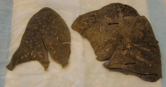
normal lung on left; bronchopneumonia (arrows) on right.
courtesy of Dr Zhaolin Xu, Department of Pathology, Dalhousie University
Treatments
- immediate
- community-acquired
- nosocomial
- nursing home
Immediate Treatments
ABCs
Oxygen
IV access
salbutamol/Ventolin 2.5mg NEB q6h
Community-acquired
Previously healthy
No antibiotics within past three months:
- macrolide (erythromycin)
- doxycyline
antibiotics with past three months:
- fluoroquinolone
- macrolide plus amoxocillin
- macrolide plus amoxocillin-clavulante
co-morbidities (COPD, diabetes, renal failure, CHF, malignancy)
no antibiotics within past three months:
- macrolide
- fluoroquinolone
antibiotics within 3 months:
- fluoroquinolone
- macrolide plus beta-lactam
suspected aspiration (oral anaerobes)
- amoxocillin-clavulanate
- clindamycin
Nosocomial Pneumonia
Anti-pseudomonal:
- ceftazedime
- piperacillin-tazobactam
further gram -ve coverage
- ciprofloxacin
- aminoglycoside (gentamicin, tobramycin) plus fluroquinolone
- aminoglycoside (gentamicin, tobramycin) plus macrolide (azithromycin)
suspect aspiration (oral anerobe coverage)
- clindamycin
- metronidazole
Nursing Home
In most patients, macrolides are good places to start. If a macrolide has been used in the past few months, a fluoroquinalone is the next line.
Guidelines for the treatment of community-acquired pneumonia are frequently updated by the Infectious Disease Society of America and the American Thoracic Society.
Consequences and Course
Complications include:
acute respiratory distress syndrome (ARDS)
pleural effusion/empyema (infection in the thoracic cavity)
lung abscess - destruction of parynchema and collection of pus
pleuritis
purulent pericarditis
hyponatremia
sepsis
brain abscess and meningitis via hematogenous spread
pyarthrosis - pus in joints
superinfection - usually drug-resistant other organisms
Increased mortality occurs in folks with:
- cardiac or pulmonary disease
- cirrhosis
- elderly
- leukopenia, leukocytosis, hypoxemia, bacteremia
The Patient
Health Care Team
Community Involvement
Resources and References
 |