
Eye
last authored:
last reviewed:
Introduction
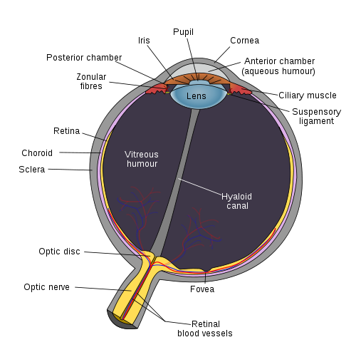
wikimedia, used with permission
- retina
- pupil
- cornea
Retina
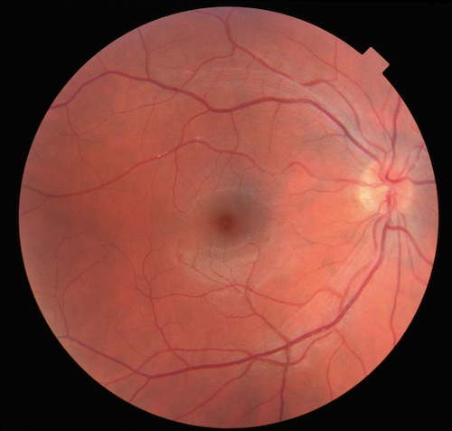
Retina, with fovea in centre and optic disc on right
The retina is set up backwards, with light passes though the nerve fibres and interneurons before striking the rods and cones.
Information leaves the rods and cones and crosses ribbon synapses onto bipolar cells, which have graded potentials. These synapse with ganglion cells that generate spikes.
There are also interneurons, and a great deal of information processing occurs even before information leaves the eye.
Ganglion cells converge on the optic disc, forming the beginnings of the optic nerve (CN II).
Rods and Cones
Rods are vey sensitive to light, operating best at night. A rod can detect a single photon, but it is very slow (perhaps 1 second).
Cones are designed for visual acuity and color perception, and are enriched in the fovea. Cones detect perhaps 100 photons but are 100 times faster.
cells are depolarized in the dark; CNG channels allowing Na and K in.
cGMP activates rhodopsin and phosphodiesterase is involved too.
in light, hyperpolarization leads to
Pupil
Pupillary Control
The sympathetic system dilates the pupil via radial iris muscles. Axons leave the cervical cord in the first thoracic root, travel around the lung, and synapse in the superior cervical gangion. Postganglionic fibres join the opthalmic branch of the trigeminal nerve near its entrance to the orbit. Damage results in Horner's syndrome.
The parasympathetic system constricts the pupil via smooth muscle in the pupillary sphincter. Fibres travel along CN III (oculomotor) from the Edinger-Westphal nucleus.
Pupillary Reflexes
Direct and Consensual Constriction
Light shone in one eye constricts both pupils. Information travels along the optic nerve (CN II), enters the superior colliculus, and synapses in the pretectal nucleus, located between the midbrain and thalamus. These cells project bilaterally to parasympathetic neurons in the Edinger-Wesphal nuclei, immediately adjacent to the CN III nucleus. Preganglionic axons travel to the ciliary ganglion, where postganglionic neurons leave to constrict pupilary smooth muscle.
Near Reflex
- lens changes shape, eyes converge, pupils constrict
- involves cortical processing, Edinger-Westphal nucleus and CN III output
Relative Afferent Pupil Defect
- picked up by swining flashlight test
- consensual response constricts a bad eye
- light shone on the bad eye dilates it
- suggests optic nerve problems
Cornea
Pupillary Control
The sympathetic system dilates the pupil via radial iris muscles. Axons leave the cervical cord in the first thoracic root, travel around the lung, and synapse in the superior cervical gangion. Postganglionic fibres join the opthalmic branch of the trigeminal nerve near its entrance to the orbit. Damage results in Horner's syndrome.
The parasympathetic system constricts the pupil via smooth muscle in the pupillary sphincter. Fibres travel along CN III (oculomotor) from the Edinger-Westphal nucleus.
Eye Movements
Types of eye movements
Congugate eye movements are those when the eyes are aligned.
fast eye movements have a velocity of 300-700 deg/sec and include saccades and the quick phase of nystagmus.
Saccades are generated in the retiucluar formation.
slow eye movements have a velocity of 20-50 deg/sec and include:
- smooth pursuit
- vestibular response
- vergence
- optokinetic
Control of Eye Movements
There are nine cardinal eye movements, controlled by 6 muscles.
CN III (oculomotor) controls the superior, inferior, and medial rectus and inferior oblique. CN IV (trochlear) controls the superior oblique, while CN VI (abducens) controls the lateral rectus.
Gaze Centres
- Lateral gaze centre is in midbrain
- eye fields are in frontal cortex
a right frontal lobe lesion causes deviation to the right
a brain stem stroke causes contralateral paralysis and eye deviation
Images are maintained on the fovea through two mechanisms - the oculomotor system, which controls our eyes, and the head movement system.
The vestibuloocular reflex is controlled by the vestibular system, which sends signals from the vestibular nucleibilaterally along the medial longitudinal fasciculus, through the PPRF (paramedian pontine reticular formation), to the oculomotor, trochlear, and abducens nuclei.
Lateral gaze is controlled by the pontine gaze centre, which sends signals from the PPRF to CN VI and CN III nuclei via the medial longitudinal fasciculus. Its disruption leads to horizontal disorder.
There are also internuclear connections among the PPRF and nuclei of CN III and VI which also involve the MLF. During lateral gaze, signals are sent from CN VI to CN III to control the medial rectus muscle. An MLF lesion will cause internuclear opthalmoplegia, leaving CN III free to work its magic during convergence but not on lateral gaze.
Pupillary Reflexes
Direct and Consensual Constriction
Light shone in one eye constricts both pupils. Information travels along the optic nerve (CN II), enters the superior colliculus, and synapses in the pretectal nucleus, located between the midbrain and thalamus. These cells project bilaterally to parasympathetic neurons in the Edinger-Wesphal nuclei, immediately adjacent to the CN III nucleus. Preganglionic axons travel to the ciliary ganglion, where postganglionic neurons leave to constrict pupilary smooth muscle.
Near Reflex
- lens changes shape, eyes converge, pupils constrict
- involves cortical processing, Edinger-Westphal nucleus and CN III output
Relative Afferent Pupil Defect
- consensual response constricts a bad eye
- light shone on the bad eye dilates it
- suggests optic nerve problems
Visual Processing
Optic nerve, chiasm, and tract
Ganglion cells coalesce at the optic disc and become myelinated forming the optic nerve. Being continuous with the CNS, the optic nerve, and in fact the sclera is covered with dura. The CNS subarachnoid space is also shared within the optic nerve, and increased intracranial pressure can be seen as papilledema.
The optic continues behind the eye until the optic chiasm. There about 50% of fibres cross. Information from the lateral retina (medial view) remains uncrossed, while that from medial retina (lateral view) crosses. In this way, information from the left side travels to the right hemisphere, and vice versa.
There are no synapses in the optic chiasm, which lies directly above the pituitary. Sinusitis or pituitary tumours can disrupt the visual stream at the chiasm.
The optic tract (or both of them) leaves the chiasm and travels around the cerebral peduncle in the midbrain to synapse within the lateral geniculate nucleus in the thalamus.
Some fibres leave the optic tract and travel to the superior colliculus in the midbrain pretectal area, where they are presumably involved in head and eye tracking.
Other fibres travel to the hypothalamus and synapse on the suprachiasmatic nucleus, providing retinal input for circadian rhythms.
Lateral Geniculate Nucleus
As information synapses within the thalamus, crossed fibres end up in one area, while uncrossed fibres go to others. This maintains retinotopy all the way through.
The LGN receives blood from two sources; problems in one vessel or the other leads to specific visual field loss.
Problems anterior of the geniculate leads to retinal atrophy and paleness; cortical damage won't lead to paleness because it is protected by the synapse.
Geniculocalcarine Pathway
Fibres leaving the LGN travel in a sheet to the occipital lobe, passing alongside and below the ventricles. Temporal fibres process the upper visual field, while fibres passing through the parietal cortex processes the lower visual field
- superior visual world is processed in the inferior cortex, and vice versa
Primary Visual Cortex (Area V1)
- dual blood supply - posterior cerebral artery and middle cerebral artery
- temporal cresent loss suggests small stroke
- visuotopy results in retinotopic map
- central region, viewed by fovea and retina just surrounding it, is responsible for about half the visual cortex
Higher Visual Processing
Distinct parts of cortex associated with specific visual functions
right = confugural
left = part-based processing
from V1, the dorsal action stream leaves to the posterior parietal cortex, while the ventral perception stream leaves to the inferotemporal cortex.
The dorsal action stream is involved with movements such as reaching.
Dorsal Action Stream
located in the posterior parietal cortex
Problems with the dorsal action stream "where" lead to:
- visual-spatial neglect - problem with attention rather than perception
- most common after right inferior parietal lesions
- Balint's syndrome
- inability to attend to and make a motor action to a location in space
- optic ataxia - cannot find objects in space
- ocular ataxia - cannot shift gaze to objects in space
- visual disorientation - cannot perceive mutiple objects sequentially
- associated with bilateral occipito-parietal problems
- motion perception (akinetopsia)
- difficulties crossing road, pouring liquids, other things having to do with movement
- problems with V5 (middle temportal gyrus)
Ventral Perception Stream
located in the inferotemporal cortex
associated with faces, objects, and words
problems with the ventral perception stream "what" lead to:
object recognition
- apperceptive agnosia
- bilateral occipital problems; likely multiple losses of visual field
- only local contour perceived
- no shape information
- cannot copy or match pictures
- associative agnosia
- unilateral or bilateral occuipital-temporal problems
- can match and copy images, though without meaning
- cannot name or sort objects
- reading can be ok or not
- appears to be a disconnect between onject and meaning
- category-specific visual agnosia
- agnosia is only for visual stimulation; touching the object will lead to recognition
- visual field defects are often associated
- face recognition (prosopagnosia)
- always posterior; mostly right, though sometimes bilateral
- sometimes colour perception loss associated
- suggests face recognition is a specific part of the brain - fusiform gyrus
- perhaps some implicit recognition of famous faces
- anti-prosopagnosia
- patients can only recognizes face, but not anything else (functionally blind)
- cerebral achromatopsia
- alexia
- loss of ability to read as a result of brain damage, but can write
- left posterior lesions often involved
- reading
- colour perception
Resources and References
 |