
Ventilation
Introduction
Ventilation is the mechanical movement of air in and out of the lungs. It is mediated by the respiratory muscles and level of lung compliance.
Distribution of Ventilation
Gas exchange occurs only in the last 5 or six airway divisions, in the transitional and respiratory zones: respiratory bronchioles, alveolar ducts, and alveolar sacs.
Va ~ VCo2 / PA CO2
The upper lung supports the weight of the rest of the lung, pulling alveoli open and decreasing compliance. This means they ventilate poorly.
The lower lung, however, bears no weight load and can therefore ventilate well.
Pleural pressure is higher at the base of the lung, which means that smallest airways are more likely to collapse.
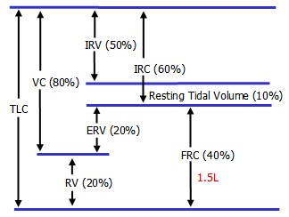
Physiologically Relevant Volumes
Dead Space
Dead space is the portion of the airways that does not allow gas exchange.
Anatomical dead space is caused by non-ventilated conducting airways, from the mouth to the terminal bronchioles.
Physiological dead space accounts for diseased areas of the lungs.
Increasing dead space will decrease alveolar ventilation:
![]()
Dead space may be estimated by the patient's weight in pounds, in ml.
Measuring Lung Volumes
Residual volume, and therefore TLC and FRC, cannot be measured directly. As such it is measured in one of two ways:
Dilution method: a volume (V1) of low solubility gas such as helium is provided and equilibirated with the person's FRC (V2). Measuring the final concentration C2, and using the equation C1 x V1 = C2 x (V1 + V2), provides V2 from which RV can be deduced (RV = FRC - ERV)
Body Plethysmograph (Body Box): a person breathing in a sealed box, with a mouthpiece and shutter, produces pressure changes when panting. Using Boyle's Law, P1V1 = P2V2, allows FRC to be measured.
Respiratory Muscles
Inspiration
The major muscle of inspiration is the diaphragm. Its downward movement
In situations of increased inspiratory need, the accessory sternocleidomastoid and scalenes can bring the ribcage up and out. Use of accessory muscles leads to their hypertrophy.
Expiration
Expiration is ususally passive, relying on the elastic properties of lungs, but forced expiration can be mediated by the intercostals and abdominal muscles. Expiratory flow limitation reduces maximum expiration velocity. These muscles can all be used to compress the abdominal contents and deflate the lungs.
- rectus abdominis
- external and internal oblique
- transversus abdominus
Factors Affecting Ventilation
- lung compliance
- airway resistance
Lung Compliance
Compliance is the change in volume divided by the change in pressure, and is the inverse of stiffness. A compliant lung has a large change in volume over a small change in pressure, signified by a steep slope. Compliance decreases as volume increases.
Compliance decreases with restrictive diseases or pulmonary edema, due to restriction of lung volume. Compliance increases with emphysema due to loss of elastic recoil. Work of breathing increases in situations with both increased and decreased compliance.
Pleural Pressure
Pleural pressure (PP) is normally - 5 as the elasticity of the lungs pulls inward while the chest wall pulls outward.
FRC, the volume of lungs after passive expiration, is the equilibrium between these two forces.
Surface Tension
The Law of LaPlace states that P = 2 T / r, with P: inside pressure, T: surface tension, and r: radius
This means that smaller alveoli contain higher inside pressure and are therefore unstable.
Surfactant, produced by Type II cells, decreases surface tension (T) which stabilizes smaller alveoli. This increases compliance and reduces opening pressure.
Work = force x distance, or pressure x change in volume.
With a slow breath, work is required to overcome the lung's elasticity, but this is returned durong expiration and no net work is done.
With rapid breath, work must both overcome lung elasticity and that caused by viscous airflow, so net work is required.
With a decrease in lung compliancy, more pressure is required to pull lung out and so work increases.
Airway Resistance
Airway resistance is determined by many parameters in the lung, both physiologic and pathologic.
Turbulent vs Laminar Flow
Re is proportional to D (diameter) V (velocity), and p (density) and inversely proportional to u (viscosity)
A low Re (below 2000) yields laminar flow, while a high Re (above 2000) is turbulent.
As the airway diameter decreases, flow switches from turbulent to laminar flow in the bronchioles. Cross-sectional area increases dramatically and velocity slows down almost to a halt.
With laminar flow, Poiseuille's Law states that the change in pressure is related to flow divided by the radius to the power of 4.
Resistance is the change in pressure divided by the flow.
As the distal airways have a much larger cross-sectional area and lower velocity, 90% of airway resistance is in the large airways. This means that small airway disease is relatively silent.
Expiratory Flow Limitation
As the lungs expand, traction on the airways increases the diameter and lowers the resistance. There is thus no limitation on inspiratory flow.
However, expiratory flow limitation occurs due to compression of small airways as pleural pressure increases and exceeds that of the airways. It is greatest at low volumes and is the cause of the effort-independent, linear segment of the flow volume loop.
Patterns of Abnormal Ventilation
hyperpnea
hyperventilation
causes of increased respiratory drive
peripheral chemoreceptor stimulation - hypoxia, hypercapnia
central chemoreceptor stimulation - hypercapnia, acidosis
Hypoventilation
Alveolar ventilation is abnormally low and always causes a raised pCO2. This results in arterial hypoxemia, unless breathing an increased O2 mixture. Hypoxemia is easily corrected by increasing inspired piO2.
Matching Ventilation and Perfusion
Ventilation and perfusion normally are regulated in relation to each other, such that there is a tight match between rates. Hypoxia causes constriction of airways, leading to rerouting of perfusion to other areas.
Both ventilation and blood flow are higher in base of lung, and VA / Q is lowest at bottom and highest at top.
VA / Q Mismatch
Mismatch can occur with a block in perfusion, ie pulmonary embolus ( VA = 0 ) or a block in ventilation (physiologic dead space; Q = 0 )
Chronic airway obstruction can lead to pulmonary hypertension and cor pulmonale.
Hypoxia
A normal A-a DO2 suggests that PO2 or alveolar ventilation is low. An abnormal A-a PO2 suggests either diffusion impairment or a VA / Q mismatch.
 |