The Respiratory System

last authored: April 2010, David LaPierre
last reviewed:


Introduction

Life is often thought of as synonymous with breathing - as the story goes, God gave Adam the "breath of life". The lungs are the cornerstone of the respiratory system, exchanging gas between the atmosphere and the blood.


Blood pumped from the heart's right ventricle enters the pulmonary arteries. Blood in the pulmonary capillaries, flows past air-filled alveoli and exchanges old carbon dioxide for fresh oxygen. This new blood flows through pulmonary veins and enters the left atrium, to be pumped through the rest of the body.


Upper Airways

Main article: upper airway

Inspired air travels through the nose and pharynx, where it is heated, humidified, and filterd of particles greater than 10 um in diameter.


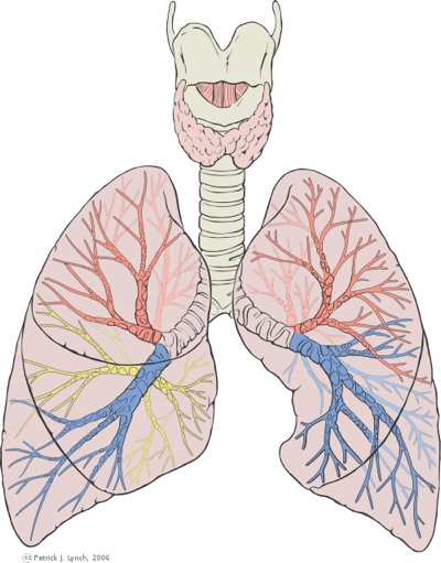
Air passes through the open larynx and through the trachea, which is 10-12 mm in diameter and held in shape by U-shaped cartilage rings.


There are perhaps 23 levels of bifurcation leading to the left and right lungs. Bronchi are the left and right initial bifurcations, and sequential bifurcations, containing cartilage. There are approximately five generations of large, intasegmental bronchi and about 15 generations of smaller intrasegmental bronchi. These become bronchioloes and end in alveoli, discussed soon.

return to top


The Lungs

Functions of the lung

  • gas exchange
  • blood reservoir
  • filter against emboli
  • metabolic location of enzymes (ie, ACE)
  • thermal reservoir
  • protecting against heat loss

The lungs are located in the thorax. The right lung has 3 lobes, 55% of total lung, while the left has two lobes. Each weighs x-x. and is x large. They are covered with the thin pleural lining - only a few cells thick. Between the lungs and chest wall is the pleural space, a very small (potential space) which contains fluid to lubricate the lungs.


Lungs are extremely elastic due to the rigid bony thorax and their cellular and connective composite.


lung landmarks

  • inferior margins: 6th (anterior)
    8th (lateral), and 10th (posteriorly) ribs
  • oblique fissures: diagonal, from T3 to the 6th rib anteriorly
  • minor fissure: horizonal line from anterior 4th rib
  • pleura is at 8,10, 12
  • major fissure goes to T3/T4 posteriorly

The majority of the lung is composed of acinar sacs, leading to a surface area of 50-100 m2 per lung. The acinar sacs contain 300 million gas-exchanging alveoli.


Between acinar sacs is the interstitium, which consists of the basement membrane of alveolar and endothelial cells - fused in the thinnest regions - and connective tissue in between. This includes:

return to top


Distal Airways

Bronchioles are bifurcations that do not contain cartilage, making them collapsible. They are also contractile due to smooth muscle. This is a principal location of asthma reactivity.


Lobules contain clusters of alveolar sacs attached to terminal bronchioles. These sacs, or acini (one acinus), are composed of respiratory bronchioles and alveoli and are the true site of ventilation.


Alveoli are gas-exchanging structures. They begin as outpouches of respiratory bronchioles but are found in greatest number in alveolar sacs.


Alveoli are in intimate contact with the endothelial cells of the lung's extensive capillary system.


The thin Type I pneumocytes make up most of the alveolar walls, while type II pneumocytes (5% if the cells) secrete surfactant to reduce surface tension and keep the alveoli inflated. Type II pneumocytes are capable of regeneration and differention into type I cells following injury.


Macrophages live in alveoli.


return to top


Pulmonary Blood Supply

The bronchial circulation nourishes the airways, and comes off the aorta.


Blood pumped from the right ventricle crosses the pulmonary valve and enters the pulmonary trunk, which divides into the two pulmonary arteries. As blood enters pulmonary capillaries, it flows past air-filled alveoli and exchanges carbon dioxide for fresh oxygen. This new blood flows along pulmonary veins and enters the left atrium.


The pulmonary vascular bed receives the entire output of the right ventricle. Because it is a low pressure system, it responds to gravity, with greatest blood flow going to the base of the lungs. Given constant alveolar pressures throughout the lung, there are three zones of pressure difference.


Zone I, at the top of the lung, has more alveolar pressure than arterial pressure. This poorly perfused area increases with physiologic dead space. The alveolus acts as a Starling resistor to prevent blood flow through poor alveoli. In zone II, in the middle, pressures are equal, and there will be moderate flow. Zone III, at the bottom of the lung flow has the greatest blood flow as arterial pressure exceeds that of the alveoli.


Blood flows well through alveoli with higher O2, and vascular resistance increases with hypoxia. In situations of generalized hypoxia, such as altitude, global vasoconstriction can result in pulmonary hypertension. Giving oxygen to patients with hypoxia decreases resistance. Acidosis and increased sympathetic tone can also cause lesser degrees of vasoconstriction.


Increasing vascular pressure causes distension of existing vessels and recruitment of new vessels, both which decrease resistance.


LV failure causes blood to back up in the lungs, causing arterial pressures to rise above alevolar pressures. This opens up apical vessels, resulting in vascular redistribution, or cephalization of blood flow.

return to top


Resources and References

return to top